hurlinghampolo.com
44
IMAGESOFPOLO.COM
revolutionary procedure was reported on
in
The Spine Journal
– an international, peer-
reviewed medical publication.
This breakthrough can be attributed to
the combined knowledge that professor
Carlstedt and I each brought from our
respective fields. As such, we are actively
and hurriedly pursuing the development
of a cross-Atlantic collaboration between the
various universities invested in researching
the advancement of spinal cord injury
rehabilitation avenues and techniques, in
order that more medical advancements can
be achieved as soon as possible.
In order for muscle function to return
after a spinal cord injury, nerve fibre
regeneration needs to take place. Currently,
damage to nerve-cell connection mechanisms
is a major impediment to regeneration, as is
the scar tissue that develops around the
supporting cells following such a trauma.
This scar tissue creates a physical barrier in
which nerve fibres get trapped and inhibits
the regeneration of the nerve cells that
connect the uninjured parts of the cord.
Most spinal cord injuries occur across
the cord, creating a disconnection between
the uninjured parts above the trauma with
those below it. This interrupts the connection
between the brain and the section of the
cord below the injury level. In other cases,
nerves attached to the cord can be torn, cut
or pulled out, causing the nerves and their
connection to the spinal cord to degenerate,
which mostly affects the nerves to the arms
or the legs, and typically results in paralysis
and loss of sensation.
During the first two weeks following
a trauma, the activity within damaged spinal
cells decreases by approximately 25 per cent,
with further deterioration over time. Swift
intervention is therefore vital to give the
afflicated person the best possible chance
of recovery. For function to be restored,
there must be a regrowth of new fibres along
the old pathways but there are cells that
inhibit this growth. The trauma to nerves
that are torn, cut or pulled out results in the
death of sensory cells. The nerve signals
diminish, which leads to abnormal sensory
signals being sent, leaving the patient with
clumsy and uncoordinated movement should
any function reappear.
In a long series of experiments in the
laboratory, Professor Carlstedt has explored
the idea of re-implanting torn muscle fibres
back into the spinal cord, and has observed
that partial muscle function returns to the
arms and legs. The recovery demonstrated
as a result of this research shows that cells
controlling nerve function from the spinal
cord can regrow within the brain and spinal
cord system – a phenomenon that has never
been observed previously. This has been
translated into a series of successful
surgeries, resulting in a return of muscle
function. The article in
The Spine Journal
described a case where a patient paralysed
below the waist due to severed nerves below
the spinal cord was, after surgery, able
to walk independently – the first report
of its kind.
Over the past 25 years, Professor
Carlstedt has worked tirelessly in pursuit
of improving the rehabilitation potentials
for spinal cord injury patients. Great
progress has been made in terms of the
improvement of motor function. However,
shortcomings in medical techniques are
proportional to delays in surgery, which is
hugely detrimental as speedy treatment of
spinal cord injury is essential in order to
delay or prevent the death of nerve cells, and
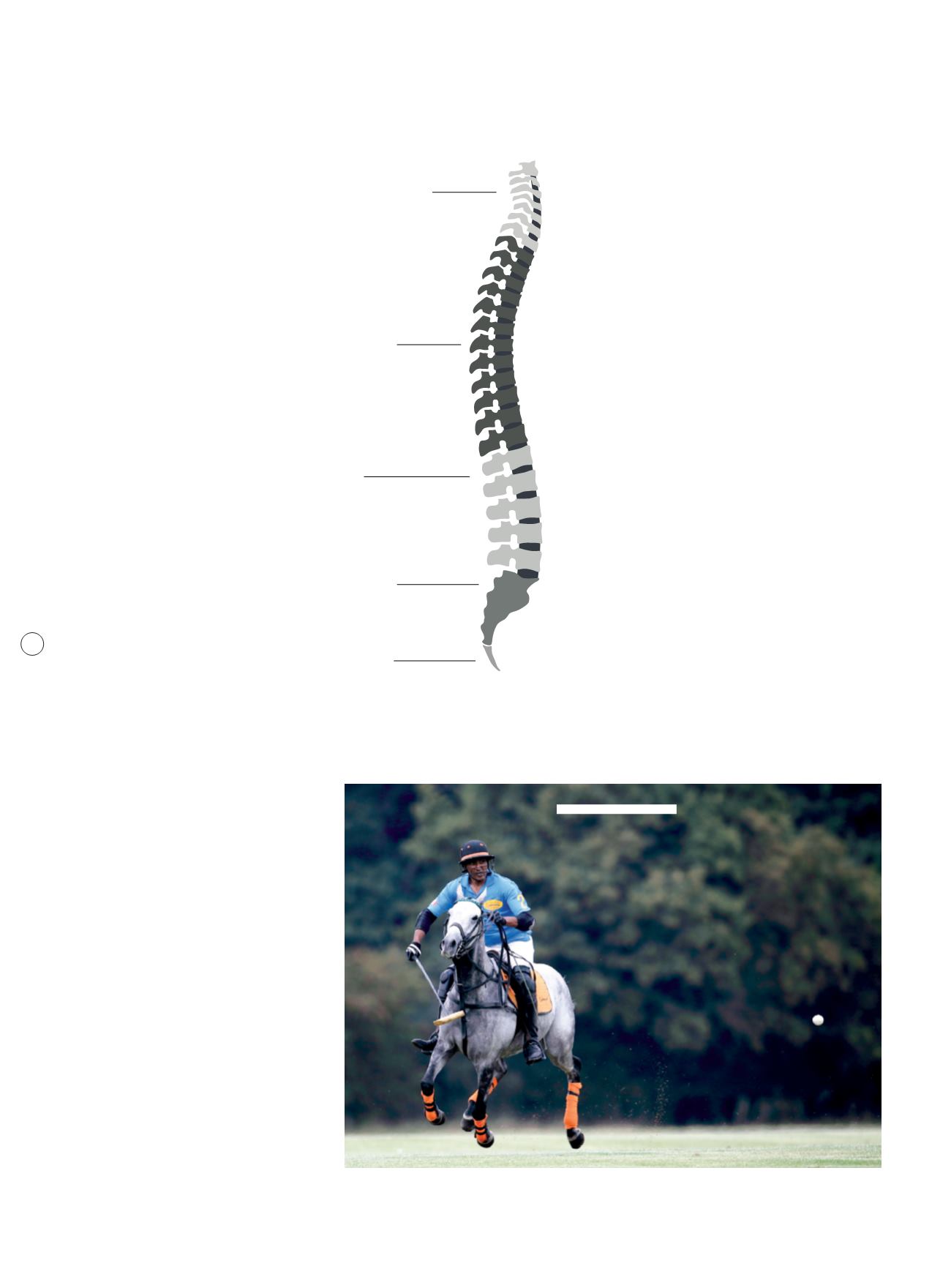
C E R V I C A L S P I N E
( 7 V E R T E B R A E )
T H O R A C I C S P I N E
( 1 2 V E R T E B R A E )
LU M B A R S P I N E
( 5 V E R T E B R A E )
S A C R U M
C O C C Y X
G R E AT P R O G R E S S H A S
B E E N M A D E I N T E R M S
O F T H E I MP R O V E ME N T O F
MO T O R F U N C T I O N


